
Medical annotation by medical imaging specialists

Morvio recruits medical imaging specialists to label medical images for AI development, or to correct the pre-annotations your model produces. They work to your protocol, and dedicated QA reviewers check their work before delivery.

We label data for AI development; we do not give clinical advice. The examples on this page are from public datasets, with each dataset's own annotations.
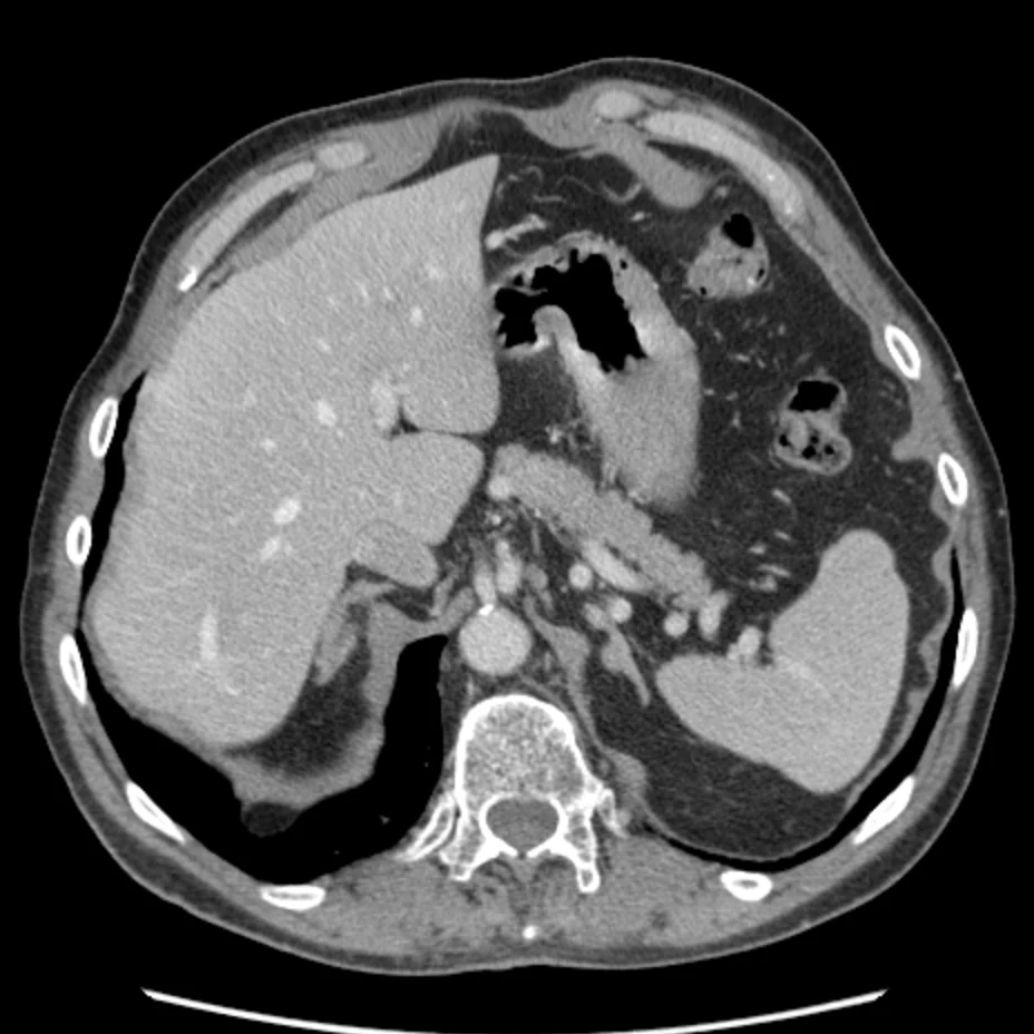
Axial abdominal CT slice from the Medical Segmentation Decathlon with one outline, the spleen, drawn in yellow; the legend names it.

CT · axial · W 400 L 40 HU · 1 of 1 class · slice 50/77 · 0.74 mm/px

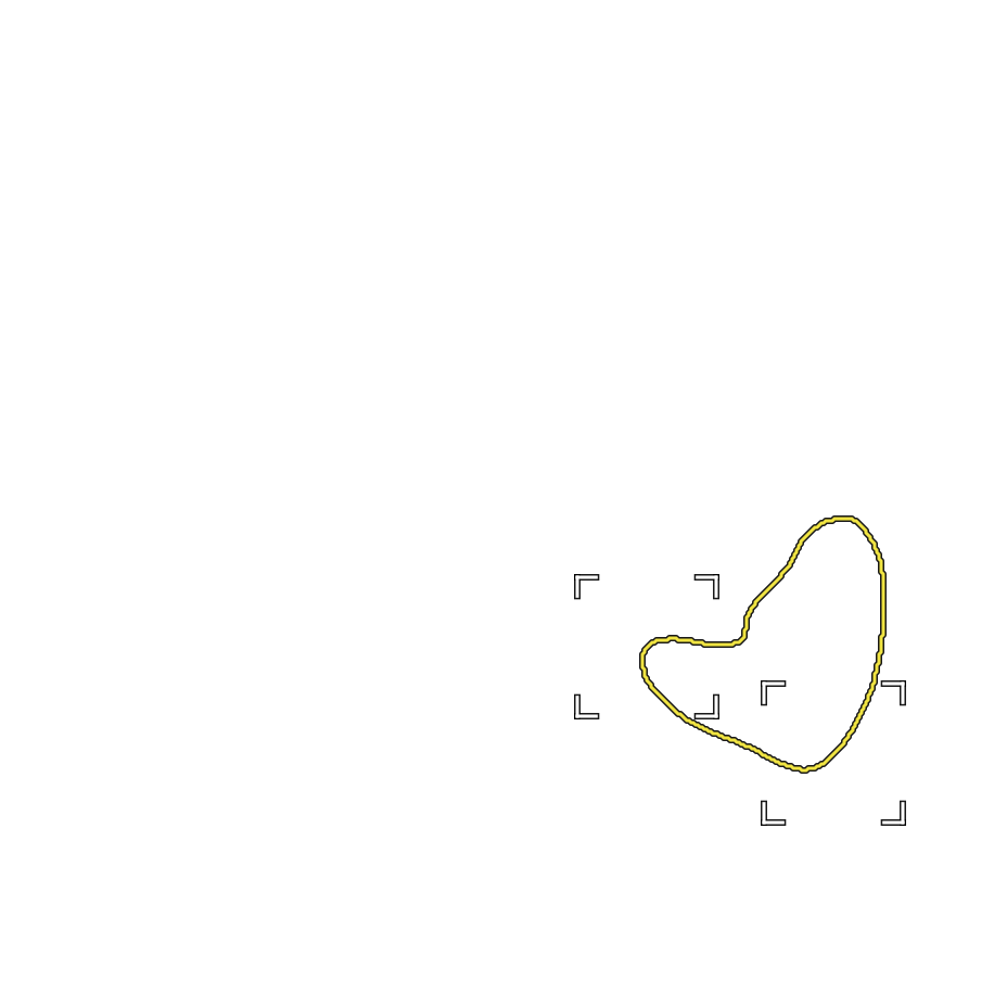
Enlarged detail, 64 by 64 source pixels without interpolation: the spleen outline over the image pixels.

Detail · 64 x 64 source px · no interpolation · 47.5 x 47.5 mm

  • spleen 1 mask
Fig. 01 · Public-dataset example · Abdominal CT · Organ segmentation (spleen)The spleen, semi-automatically segmented and manually adjusted by an expert abdominal radiologist, on one axial CT slice.Image and mask: Medical Segmentation Decathlon, Task09_Spleen (Simpson et al., 2019; Antonelli et al., 2021), CC BY-SA 4.0; the spleen was segmented semi-automatically and the contours manually adjusted by an expert abdominal radiologist; one axial slice, cropped to the body, display window W 400 L 40 HU, annotation unchanged. This figure is shared under CC BY-SA 4.0. Credits
Price basis
Per image or per expert hour
After the pilot, we confirm the production price in writing.
Who does the work
Medical imaging specialists
Each specialist's registration is checked in their country of practice.
Review
Dedicated QA reviewers
They check the specialists' work before delivery.
How to start
Send a project brief
Describe the task in five short steps. You do not need to send any data.

What the specialists do

On a typical project, a specialist opens each image and checks every box, outline or label against your protocol. They accept it, adjust it or flag it. Cases outside the protocol go to you, through the project lead.

The example images on this page come from public datasets. The annotations are each dataset's own, drawn unchanged; Morvio did not create or review them. Credits
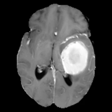
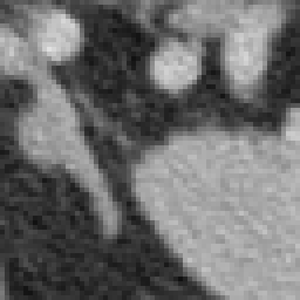
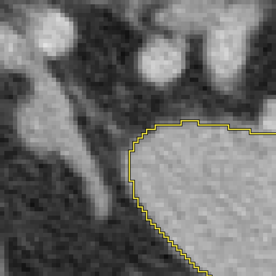
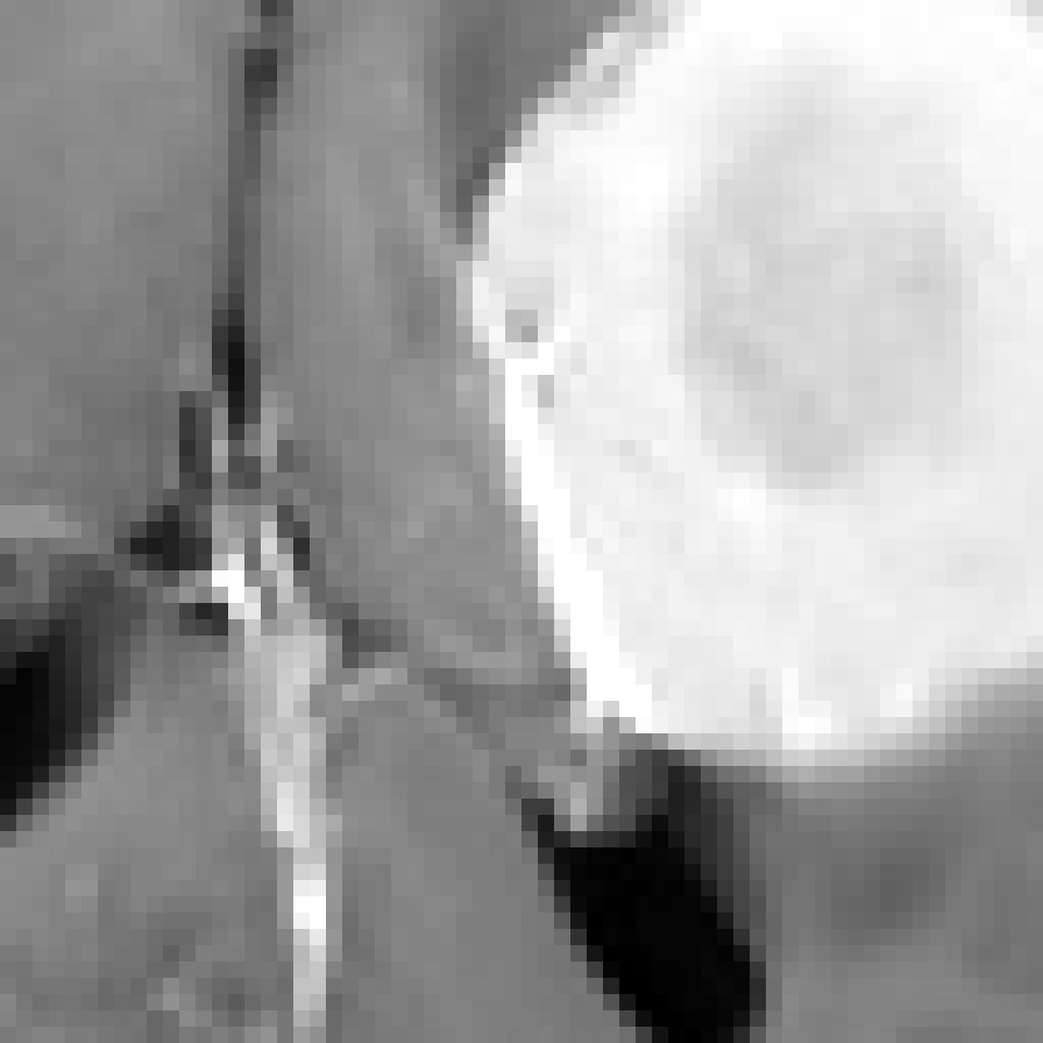
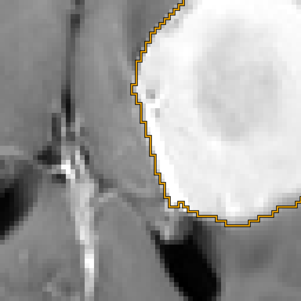
Axial brain MRI slice (T1gd) from the Medical Segmentation Decathlon with the outline of the dataset's enhancing-tumour label, one region.

MRI · T1gd · axial · slice 68 of 155 · 1 of 3 label classes shown · display 290–1207 · 1 mm/px

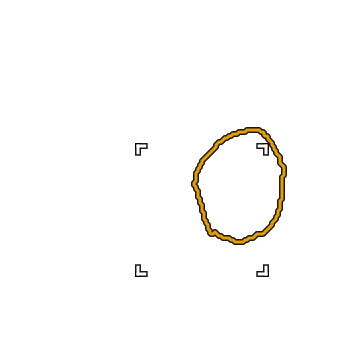
Enlarged detail, 64 by 64 source pixels without interpolation: the enhancing-tumour outline over the image pixels.

Detail · 64 x 64 source px · 64 x 64 mm · no interpolation

  • enhancing tumour 1 region
Fig. 02 · Public-dataset example · Brain MRI · Enhancing tumour segmentationThe enhancing tumour labelled by the dataset on one T1gd slice. The dataset's edema and non-enhancing tumor labels are not drawn: they are defined on other MRI sequences and are not visible on this one.Image and labels: Medical Segmentation Decathlon, Task01_BrainTumour, from BraTS (Simpson et al., 2019; Bakas et al., 2017), CC BY-SA 4.0; annotations approved by expert board-certified neuroradiologists; one slice, cropped, linear display window 290 to 1207, enlarged 2×, annotation unchanged. This figure is shared under CC BY-SA 4.0. Credits
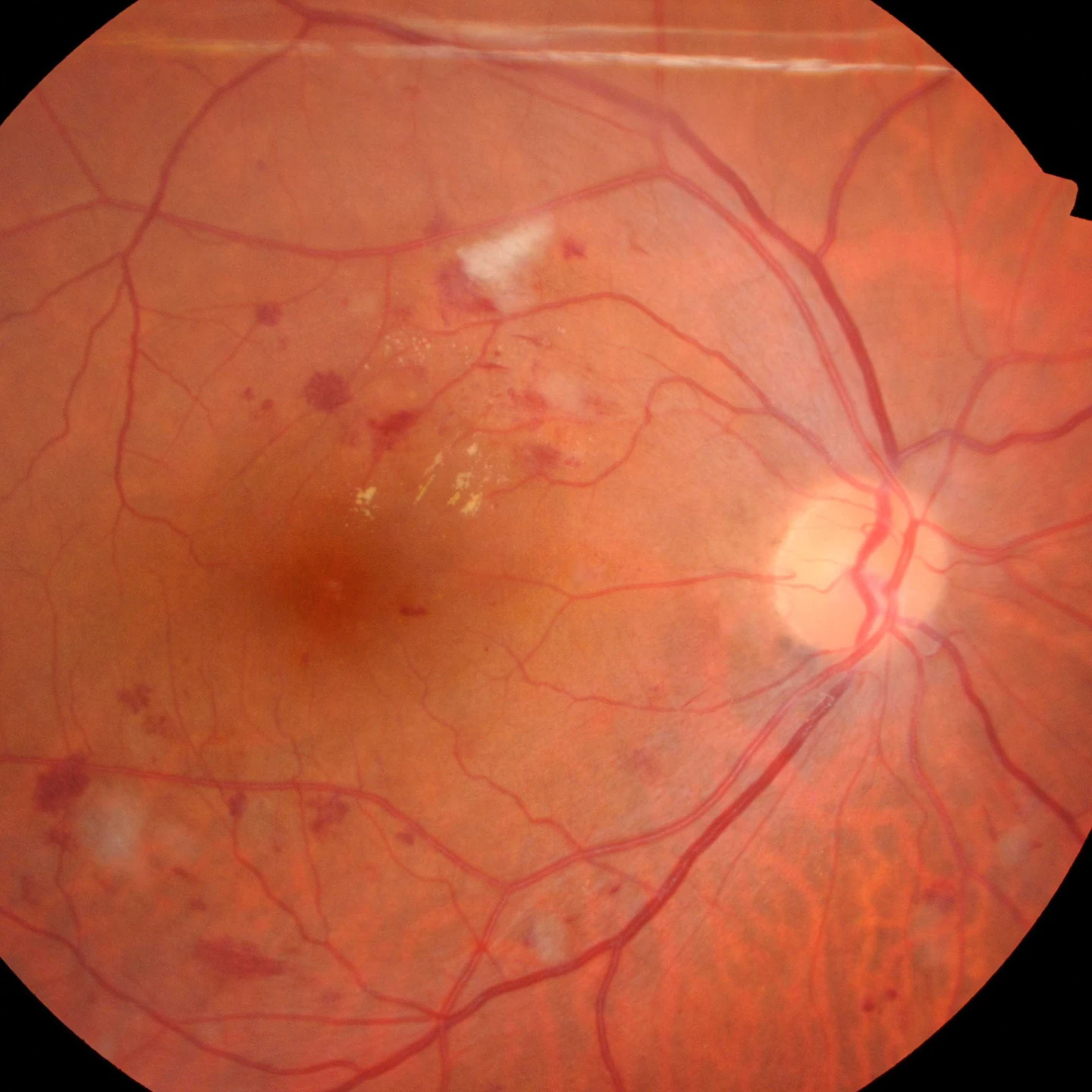
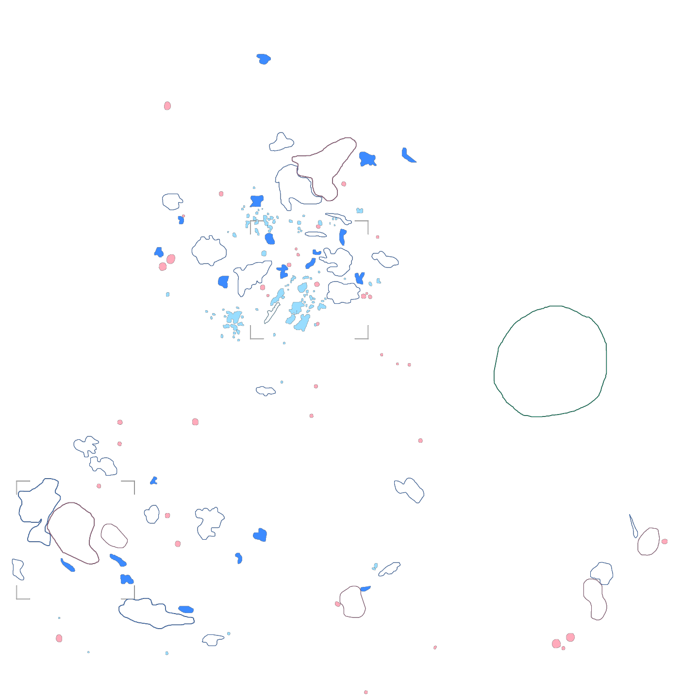
Colour fundus photograph from IDRiD with the dataset's pixel masks: 93 hard exudate, 44 haemorrhage, 38 microaneurysm and 6 soft exudate regions and the optic disc.

Colour fundus photograph · Kowa VX-10α, 50° field · pixel masks · 5 classes · 182 regions

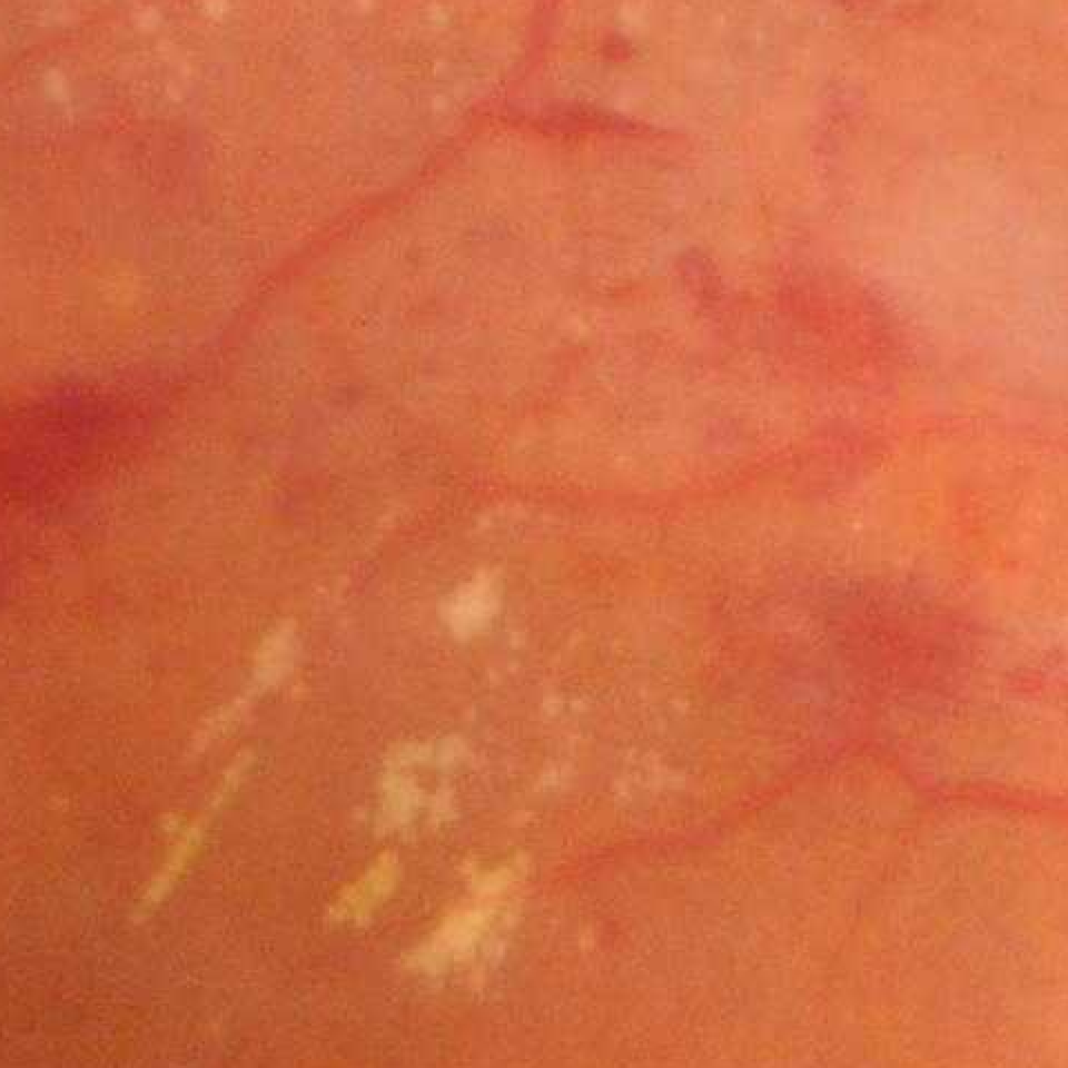
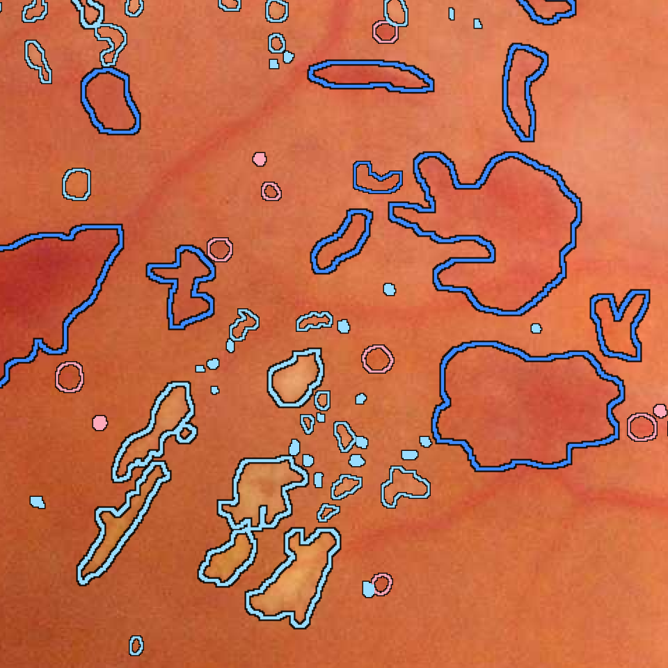
Enlarged detail, 480 by 480 source pixels without interpolation: lesion mask outlines from the dataset over the photograph's pixels.

Detail · 480 x 480 source px · no interpolation

  • Microaneurysms 38 regions
  • Haemorrhages 44 regions
  • Hard Exudates 93 regions
  • Soft Exudates 6 regions
  • Optic Disc 1 region
Fig. 03 · Public-dataset example · Colour fundus photograph · Lesion and optic disc segmentationPixel-level masks of microaneurysms, haemorrhages, hard exudates and soft exudates, and of the optic disc, as provided in IDRiD for this image.Image and masks: IDRiD (Porwal et al., 2018), CC BY 4.0; pixel-level annotation by a master's student, reviewed by two retinal specialists and finalised at consensus; square crop, reduced to 1440 px, details pixel for pixel, annotation unchanged. Credits
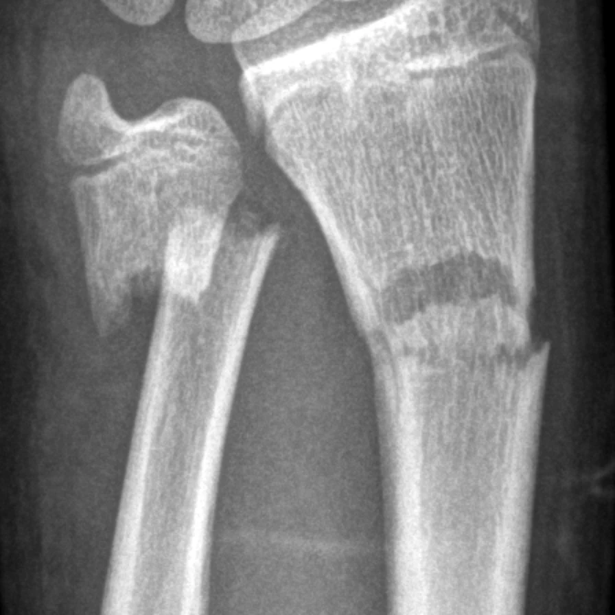
Wrist radiograph, AP projection, from the GRAZPEDWRI-DX dataset, with the dataset's 2 bounding boxes of the class fracture drawn in yellow.

X-ray · wrist · AP projection · bounding boxes · 2 fracture · 0.144 mm/px · display 0.5–99.5 percentile

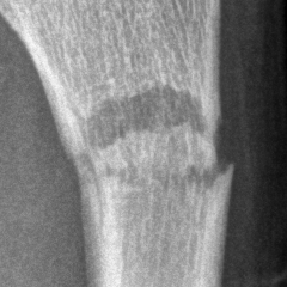
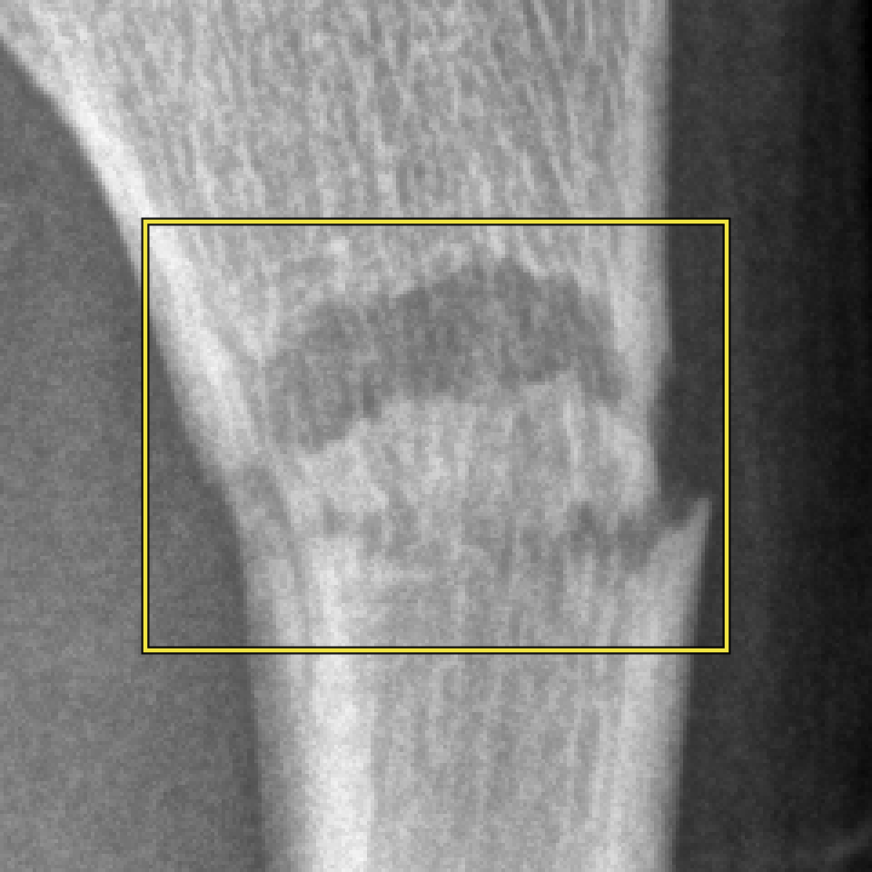
Enlarged detail, 240 by 240 source pixels without interpolation: one fracture bounding box over the image pixels.

Detail · 240 × 240 source px · 34.6 × 34.6 mm · no interpolation

  • fracture 2 boxes
Fig. 04 · Public-dataset example · Wrist radiograph · Fracture localisation with bounding boxesBounding boxes of the class “fracture” on a wrist radiograph in AP projection, as annotated in the dataset.Image and boxes: GRAZPEDWRI-DX (Nagy et al., 2022), CC BY 4.0; annotations validated by board-certified paediatric radiologists; cropped, one linear display window, enlarged 2×, annotation unchanged. Credits

Task types

Pre-annotation correction
Review and correction of boxes, outlines and labels produced by your model
Segmentation
Outlines of organs, structures and findings
Classification
Image-level or study-level labels
Signal annotation
Beat and rhythm labels on ECG strips
Escalation
Cases outside the protocol flagged and routed, never guessed
More examples: histopathology, ultrasound, ECG and dental X-ray
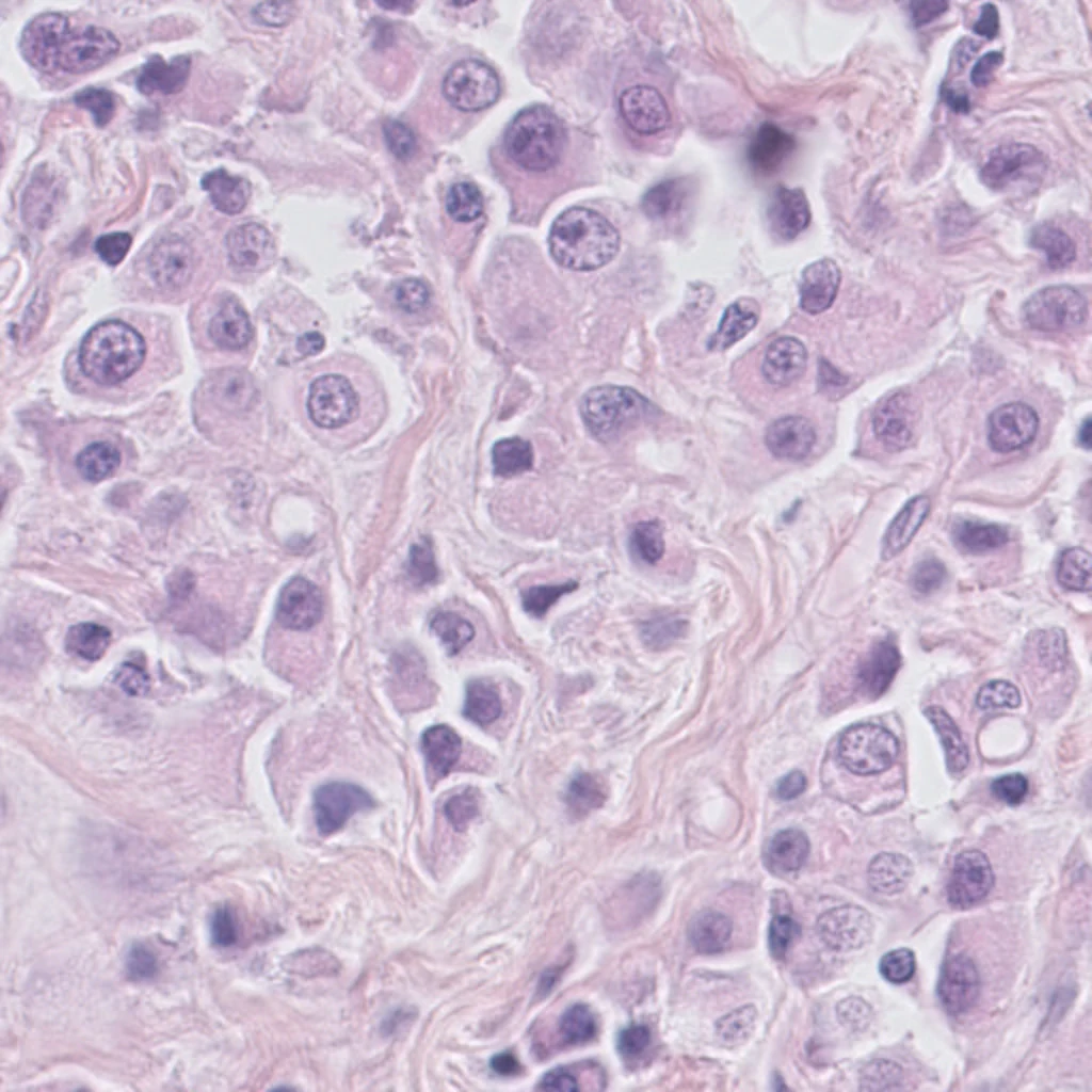
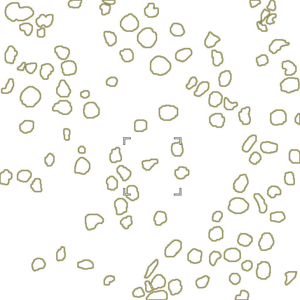
H&E-stained tissue tile from the TNBC dataset with outlines of the 103 nucleus regions in the dataset's mask; the legend gives the count.

H&E · 40× (dataset authors) · pixel masks · 103 nucleus regions (8-connected)

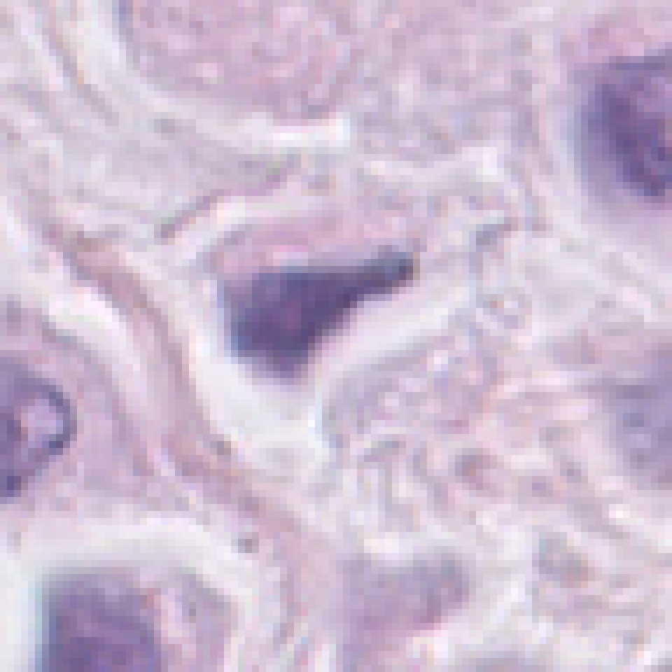
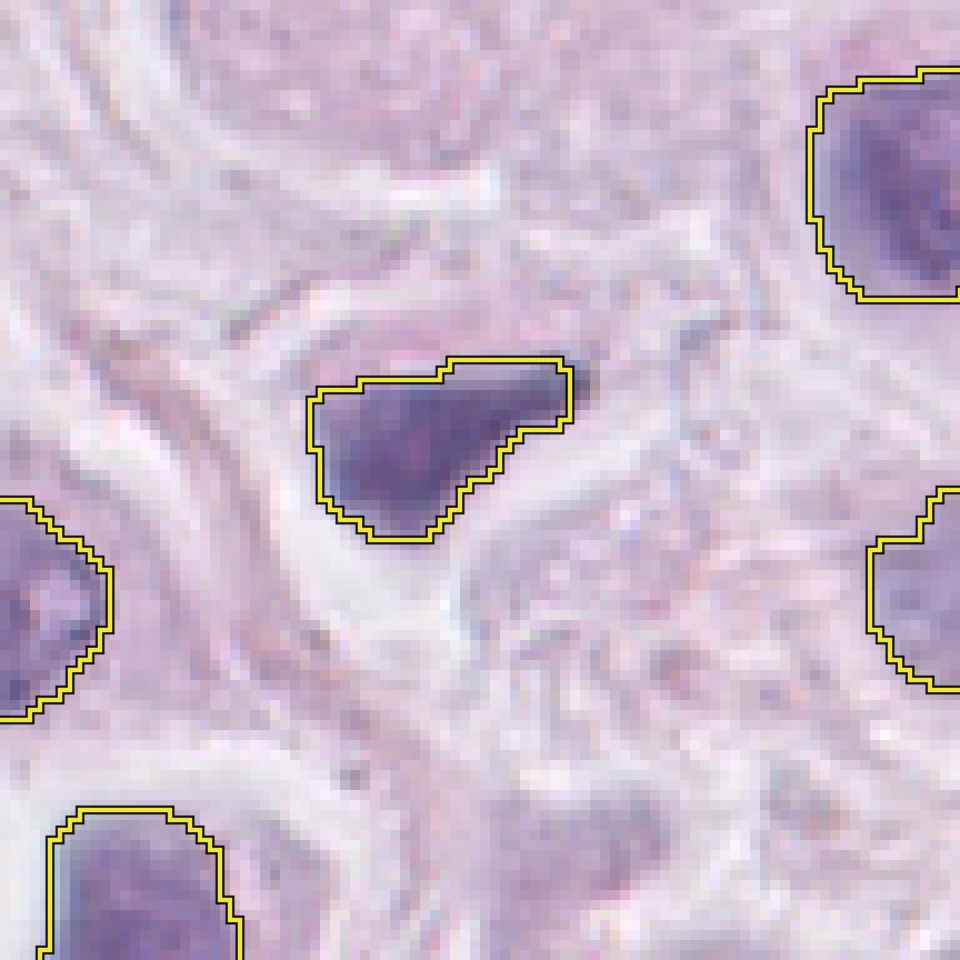
Enlarged detail, 96 by 96 source pixels without interpolation: nucleus outlines from the dataset's mask over the image pixels.

Detail · 96 x 96 source px · no interpolation

  • nucleus 103 regions
Fig. 05 · Public-dataset example · Histopathology, H&E · Nucleus segmentationNuclei annotated at pixel level in the dataset's binary mask for this 512 x 512 tile, every region outlined.Image and masks: TNBC nucleus segmentation dataset (Naylor et al., 2019), CC BY 4.0; masks as published by the dataset; whole tile, enlarged 2×, annotation unchanged. Credits
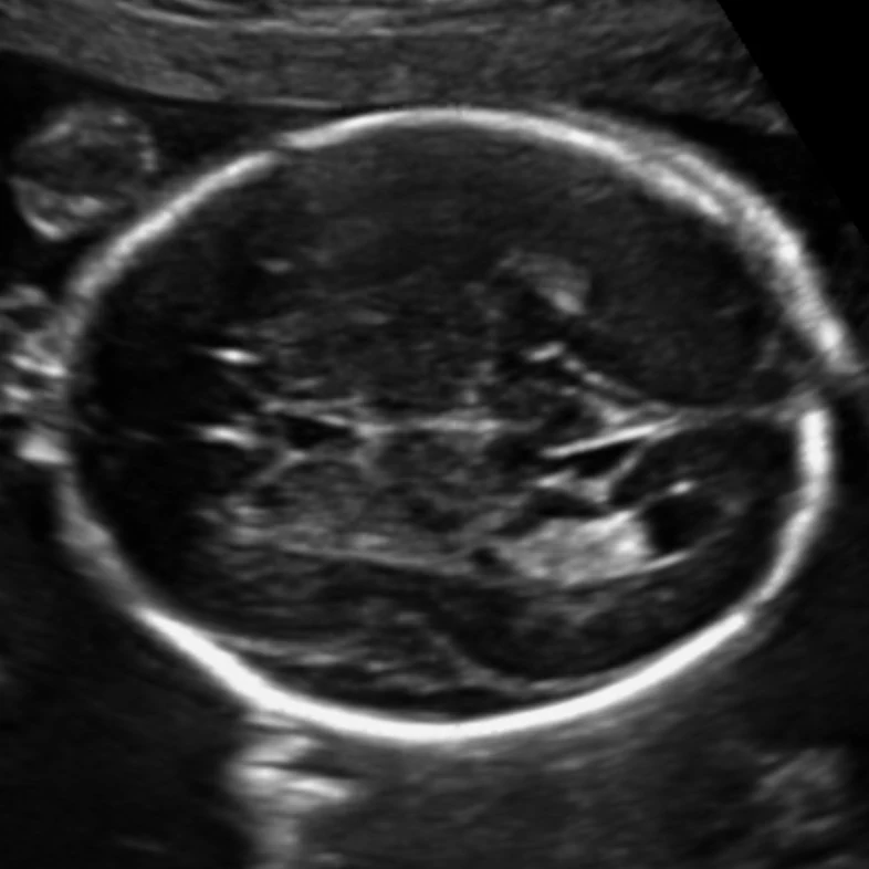
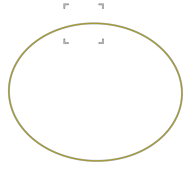
Fetal head ultrasound image from the HC18 dataset with one ellipse, the sonographer's head-circumference annotation, drawn in yellow.

Ultrasound · fetal head · ellipse · HC 204.6 mm · 0.202 mm/px

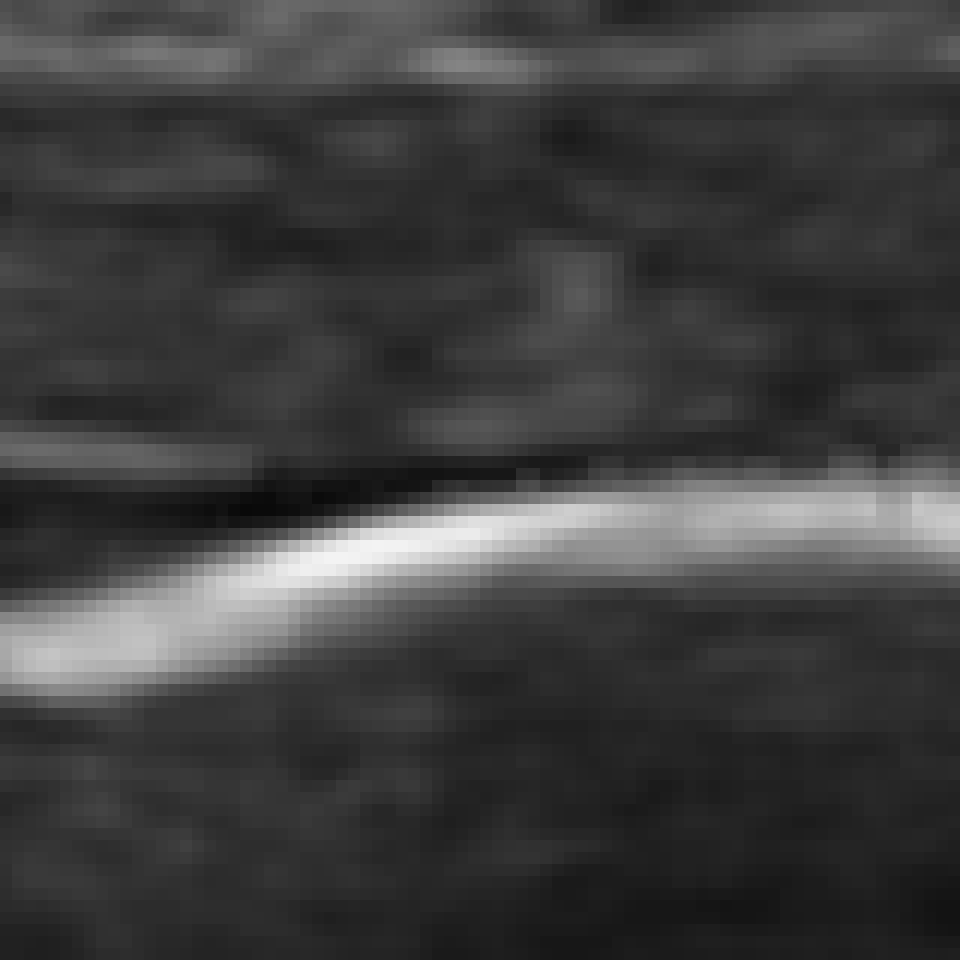
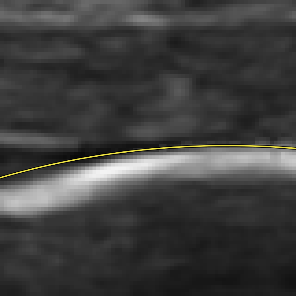
Enlarged detail, 80 by 80 source pixels without interpolation: the head-circumference ellipse over the skull echo.

Detail · 80 x 80 source px · no interpolation · 16.2 x 16.2 mm

  • head circumference 1 ellipse
Fig. 06 · Public-dataset example · Fetal ultrasound · Ellipse measurement (head circumference)Head circumference annotated by the sonographer as an ellipse that best fits the circumference of the head; the dataset records it as 204.6 mm. The dataset paper notes that a drawn ellipse never fits the outer edge of the skull perfectly.Image and ellipse: HC18 (van den Heuvel et al., 2018), CC BY 4.0; the ellipse was drawn manually by the sonographer during the exam; cropped, shown as distributed, ellipse fitted to the dataset's annotation line, unchanged. Credits
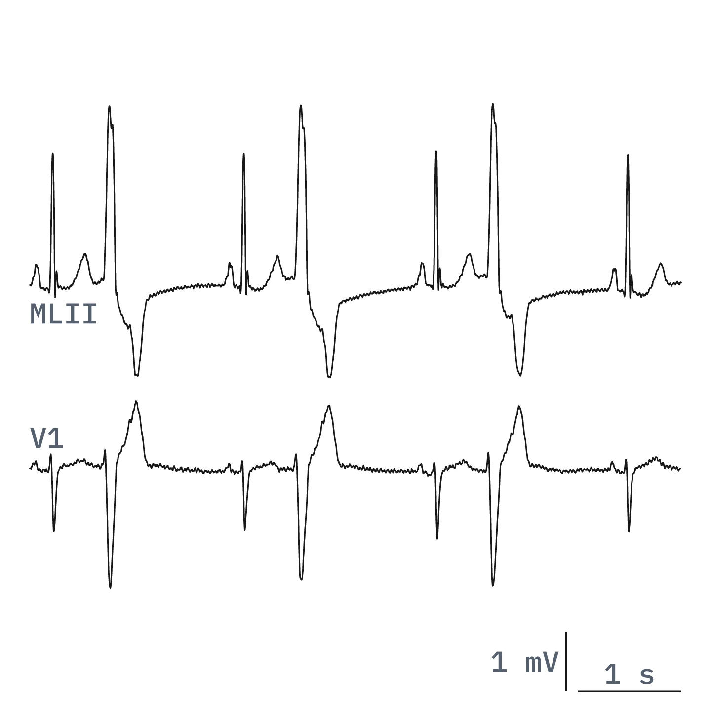
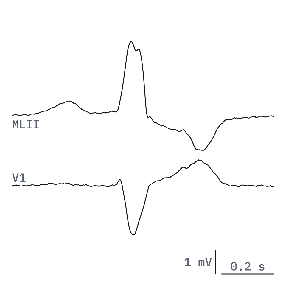
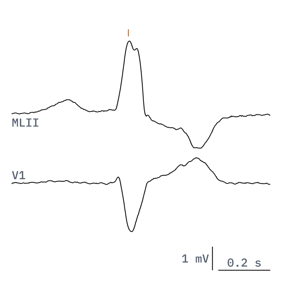
Two-lead ECG strip (MLII and V1) from MIT-BIH record 119 with the database's labels for 7 beats (4 normal, 3 premature ventricular) and two rhythm changes.

ECG · MLII, V1 · 360 Hz · beat and rhythm labels · 7 beats (4 N, 3 V) · record 119, 7:45.27-7:51.58

Enlarged one-second detail of both leads around the first premature ventricular contraction, with its beat label and the rhythm label ventricular bigeminy.

Detail · 1.00 s · samples 167612-167971

  • Normal beat (N) 4 beats
  • Premature ventricular contraction (V) 3 beats
  • (B · Ventricular bigeminy 1 rhythm label
  • (N · Normal sinus rhythm 1 in window, 1 in force at start
Fig. 07 · Public-dataset example · ECG · Beat and rhythm labelsEach beat labelled with the database's beat code and each rhythm change with its rhythm label, at the stored sample.Signal and labels: MIT-BIH Arrhythmia Database (Moody and Mark, 2005), ODC-By 1.0; reference labels from two cardiologists' independent review, with disagreements resolved by consensus; 6.31 s window plotted from the stored samples without filtering, annotation unchanged. Credits

Dental

Dental radiographs: each tooth outlined and given its FDI number, and other dental labels to your protocol.
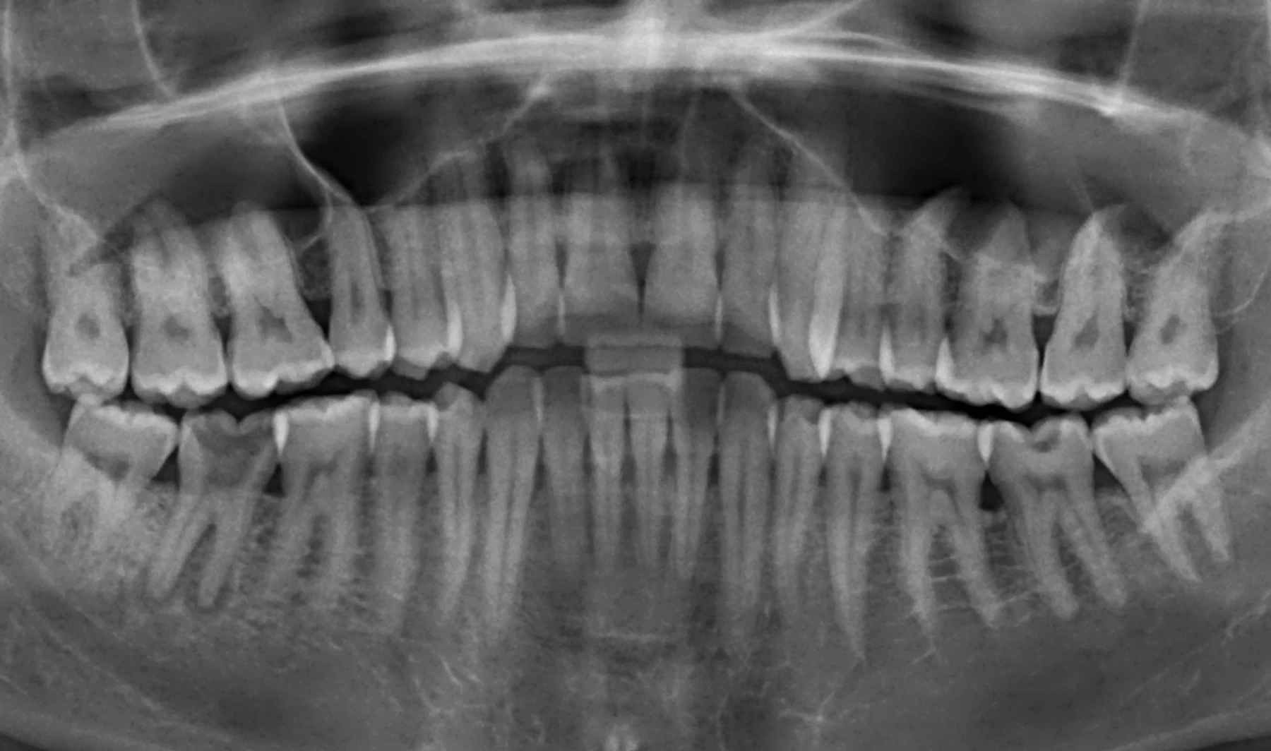
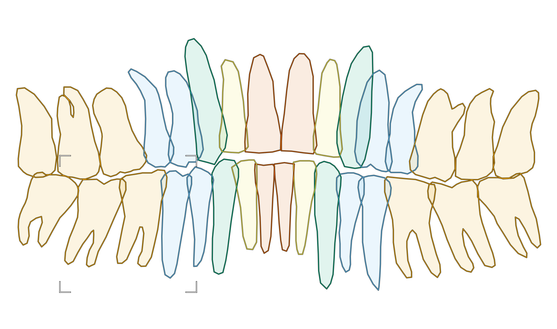
Panoramic dental radiograph with 32 tooth outlines from the OPG teeth segmentation and numbering dataset, coloured by tooth class and numbered FDI 11 to 48.

Panoramic radiograph · instance polygons · 32 teeth · FDI numbers · 8-bit as distributed

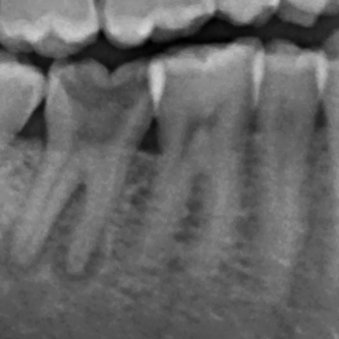
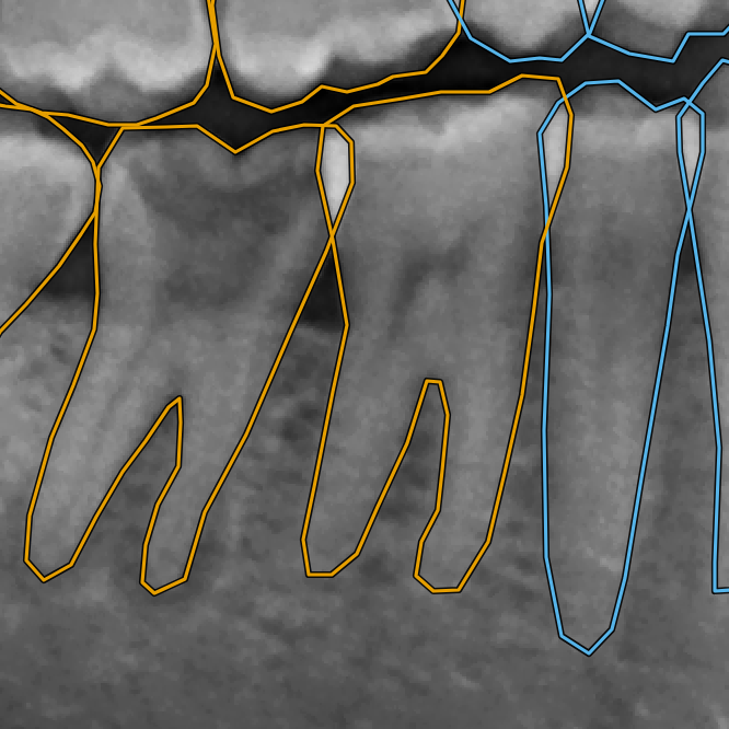
Enlarged detail, 333 by 333 source pixels without interpolation: outlines of teeth 45, 46 and 47, and parts of neighbouring teeth, over the image pixels.

Detail · 333 x 333 source px · no interpolation

  • molar 12 teeth
  • premolar 8 teeth
  • canine 4 teeth
  • lateral incisor 4 teeth
  • central incisor 4 teeth
Fig. 08 · Public-dataset example · Panoramic radiograph · Instance polygons with FDI numbersEvery tooth outlined as its own polygon and labelled with its tooth class and FDI number, as in the dataset.Image, outlines and numbers: OPG teeth segmentation and numbering dataset (Adnan and Umer, 2024), CC BY 4.0; annotations by a specialist dentist, verified by another; cropped, image as distributed, annotation unchanged. Credits

Indicative price

Per image or per expert hour.

The price depends on:

  • the modality;
  • the number of labels per image;
  • whether you supply pre-annotations;
  • review depth.

After the pilot, we confirm the production price in writing.

Get an estimate

Specification

Protocol
Your labelling protocol, or one we draft with you
Tools
Your tool, or a widely used open-source one

Who does the work

Medical imaging specialists, matched to the modality and the task. Depending on the project, the team can include radiographers or radiologists.

  • Each specialist's registration is checked in their country of practice before they work on your data.
  • Before work starts, you see each specialist's country of practice, registration body and years of practice, with their consent.
  • Image types: X-ray, CT, MRI, ultrasound

Your project team

Like every Morvio project, a medical project has its own team: a team leader, specialists recruited for your modality, and dedicated QA reviewers. The team meets daily, and everyone signs an NDA before seeing data. Your project lead is the founder, Khaled Zerouki.

Quality method

How we vet medical specialists

  1. 1

    Registration check

  2. 2

    Domain task

  3. 3

    Protocol calibration

  4. 4

    Reviewed first batch

  5. 5

    Ongoing checks

Questions

Do your specialists diagnose patients?

No. We label data for AI development; we do not give clinical advice. Specialists label images against your protocol only.

Can you correct our model's pre-annotations instead of labelling from scratch?

Yes. Specialists accept, adjust or flag each pre-annotation against your protocol.

Can we see who will label our images?

Yes. Before work starts, you see each specialist's country of practice, registration body and years of practice, with their consent.

Which image types do you cover?

X-ray, CT, MRI and ultrasound. For another modality, tell us in the estimate form. We confirm it only once vetted specialists are in place.

Do the images have to leave our environment?

No. We can work inside your tool. If we use an open-source tool, we agree where it is hosted before any image moves.

Are you a medical imaging specialist? We recruit per project and pay for the work.

Apply to join

Discuss your project

Two ways to start. Both reach Khaled Zerouki.

Book a call. Pick a time in your own time zone.

Book a call

Scope a pilot. A fixed-price batch of your real task, with acceptance criteria agreed in writing.

Scope a pilot

Want a price first?

Get an estimate

We reply within 1 business day.¹ NDA on request. We can plan and price your project without seeing your data.

¹ Business days are Monday to Friday, UK time, excluding public holidays in England and Wales.